")
")
A 64-year old female presented with fever, diffuse muscle pains and extensive areas of erythema and edema over her thighs and arms. She reported that these skin lesions developed over a few days. In addition the patient noticed generalized edema.
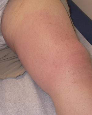
On examination, she looked acutely ill. She could barely walk with the support of two of her family members. Generalized edema was evident. The patient weighed 100 kg (her usual weight was 75 kg). Her blood pressure was normal but she was tachycardic (104 beats per minute) and tachypneic (20 respirations per minute) with oxygen saturation level on air of 95%. Her temperature was 390C. Areas of erythema were noted over her right and left thigh (Figure) and her arms extending to the anterior chest wall and breasts. These painful skin lesions were accompanied by warmth and tenderness. An area of fluctuance could be felt in her right thigh. On auscultation of the chest there were decreased breath sounds bilaterally. The rest of the physical examination was normal.
A hematocrit of 23.7% (hemoglobin 7.6gr/dl) accompanied by leukocytosis (19650/mm3 with 87.4% neutrophils with a shift to left) and mild thrombocytosis (501000/mm3) were present. C-reactive protein levels were increased at 26.71 mg/dl (normal up to 0.5) and erythrocyte sedimentation rate was 130 mm/1st hour (normal 0-20). There was a striking increase in creatine phosphokinase with serum levels of 6763 IU/l [upper limit of normal (ULN)=215 IU/l] accompanied by an increase in lactic dehydrogenase serum levels (LDH: 302 IU/l, ULN=190) and serum transaminases levels [AST: 230 IU/l (ULN=37) and ALT: 129 IU/l (ULN=65)]. Urine dipstick was positive for hemoglobin (++). A significant hypoalbuminemia was present [1.7 gr/dl (normal >3.5]. On urine microscopy there were 4-5 leukocytes per high power optical field, while there were no erythrocytes. Her electrocardiogram was normal. Chest X-rays revealed moderate bilateral pleural effusions.
What is the diagnosis?
 Matthew E. Falagas, MD, MSc, DSc
 George Peppas, MD, PhD
 Panayiotis J. Papagelopoulos, MD, DSc
 Konstantinos Rellos, MD